Skeletal Muscle Striations: What's the Secret Code?

The sarcomere, a fundamental unit within skeletal muscle, exhibits a distinct banding pattern critical to muscle function. These bands, responsible for the visible striations of skeletal muscle, are not random; the striations of skeletal muscle are produced by specific arrangements of proteins. Actin and myosin, the primary protein filaments, interact dynamically during muscle contraction. Electron microscopy has allowed researchers to visualize these arrangements in detail, revealing how the striations of skeletal muscle are produced by the intricate interplay of these molecular components within the sarcomere, ultimately influenced by factors such as calcium concentration and the signaling pathways discovered by pioneers like Andrew Huxley.
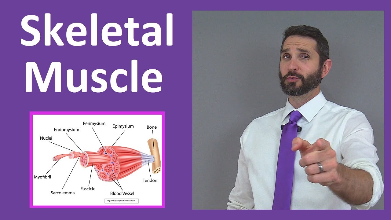
Image taken from the YouTube channel RegisteredNurseRN , from the video titled Skeletal Muscle Tissue: Contraction, Sarcomere, Myofibril Anatomy Myology .
Unlocking the Code of Skeletal Muscle Striations
Skeletal muscle, the engine of our movement, empowers everything from a delicate blink to a powerful sprint. Its function is vital to our interaction with the world. Without it, even the simplest actions would be impossible.
But what governs the incredible power and precision of these muscles?
Did you know that the seemingly uniform tissue of skeletal muscle is actually a highly ordered landscape? It's marked by a repeating pattern of light and dark bands known as striations, almost like tiny barcodes that dictate how our muscles work.
These striations aren't just a visual curiosity; they're the key to understanding how our muscles generate force.
At the heart of skeletal muscle organization lies the sarcomere, a repeating unit whose protein arrangement is directly responsible for the striated appearance. Ultimately, the striations are a direct consequence of the sarcomere's organization.
They are also fundamental to the process of muscle contraction itself.
In essence, these patterns unlock the secrets to how our muscles move us.
The Essence of Skeletal Muscle
Skeletal muscle is a type of striated muscle tissue that is attached to bones and responsible for voluntary movements. It constitutes a significant portion of our body mass and plays a crucial role in maintaining posture, locomotion, and overall physical function.
It allows us to interact with our environment.
Its importance extends far beyond simple movements. Skeletal muscle contributes to temperature regulation, nutrient storage, and even respiratory function.
The health and proper functioning of skeletal muscle are therefore paramount to our overall well-being.
The Intriguing World of Striations
What creates the striking, banded appearance of skeletal muscle? The answer lies in the highly organized arrangement of proteins within the muscle fibers.
These repeating units, or striations, are not random. They are a direct reflection of the underlying structure responsible for muscle contraction.
Think of them as the visual manifestation of a complex molecular machine.
These bands are caused by the arrangement of contractile proteins. They are actin and myosin, within the sarcomere.
The striations are key to understanding muscle function.
Thesis: Striations as the Key to Muscle Organization and Contraction
The presence of striations in skeletal muscle is not merely a superficial characteristic. It’s a fundamental aspect of its organization and function.
The striations are a direct result of the highly ordered arrangement of proteins within the sarcomere, the basic contractile unit of muscle.
This arrangement is not arbitrary; it's precisely orchestrated to enable the sliding filament mechanism, the process by which muscles generate force and contract.
Therefore, striations serve as a visual roadmap to understanding the intricate relationship between muscle structure and function. It also highlights how sarcomere protein arrangement is crucial for muscle contraction.
Skeletal muscle's remarkable capabilities are rooted in its distinct structural elements. Understanding the organization of these elements is key to understanding muscle function. Delving deeper, we find that the striations observed in skeletal muscle are not arbitrary patterns. Instead, they reflect a highly organized internal architecture. This architecture is defined by the sarcomere.
The Sarcomere: The Fundamental Building Block
The sarcomere is the basic contractile unit of skeletal muscle. It is the smallest functional unit responsible for muscle contraction. Imagine it as a single link in a chain, where many sarcomeres are linked together in series to form a myofibril.
Each sarcomere is delineated by distinct components, each with a specific role in the contraction process.
Key Components of the Sarcomere
The sarcomere's structure is defined by the arrangement of several key protein filaments and zones. These include actin, myosin, the Z-disc, I-band, A-band, H-zone, and M-line. Let's explore each of these in detail:
Actin: The Thin Filament
Actin filaments are the thin filaments within the sarcomere. These filaments are composed of globular actin (G-actin) monomers that polymerize to form long, helical strands of filamentous actin (F-actin).
Think of them as twisted ropes, providing a track for the myosin filaments to grab onto.
Associated with the actin filaments are two other proteins: tropomyosin and troponin. These regulatory proteins play a crucial role in controlling muscle contraction, which will be discussed in greater detail later.
Myosin: The Thick Filament
Myosin filaments are the thick filaments of the sarcomere. These filaments are composed of myosin protein molecules.
Each myosin molecule has a head and a tail region. The tail regions intertwine to form the shaft of the thick filament, while the globular heads project outwards.
These heads are crucial because they bind to actin and generate the force that drives muscle contraction.
Z-Disc (or Z-Line): The Sarcomere's Boundaries
The Z-disc, also referred to as the Z-line, marks the boundaries of each sarcomere. It serves as an anchor point for the actin filaments.
Imagine the Z-disc as the end of a train track, where the actin filaments are securely attached. The Z-disc not only defines the sarcomere's length but also provides structural support.
I-Band: The Region of Actin Alone
The I-band is the region of the sarcomere that contains only actin filaments. It appears as a lighter band under a microscope because it lacks the thicker myosin filaments.
The I-band spans two adjacent sarcomeres and is bisected by the Z-disc. During muscle contraction, the I-band narrows as the actin filaments slide past the myosin filaments.
A-Band: The Region of Myosin and Actin Overlap
The A-band is the region of the sarcomere that contains myosin filaments, along with overlapping actin filaments. It is the darkest band when viewed under a microscope due to the presence of both thick and thin filaments.
The length of the A-band remains relatively constant during muscle contraction. It represents the length of the myosin filament.
H-Zone: The Region of Myosin Alone
The H-zone is the region within the A-band that contains only myosin filaments. It appears as a lighter region within the darker A-band.
During muscle contraction, the H-zone narrows as the actin filaments slide towards the center of the sarcomere, increasing the overlap between actin and myosin.
M-Line: The Myosin Anchor
The M-line is a dark line in the middle of the H-zone. It is formed by proteins that connect and align the myosin filaments.
The M-line helps to maintain the structural organization of the sarcomere, ensuring that the myosin filaments are properly positioned.
Skeletal muscle's remarkable capabilities are rooted in its distinct structural elements. Understanding the organization of these elements is key to understanding muscle function. Delving deeper, we find that the striations observed in skeletal muscle are not arbitrary patterns. Instead, they reflect a highly organized internal architecture. This architecture is defined by the sarcomere.
Decoding the Striation Pattern: A Visual Guide
Having dissected the individual components of the sarcomere, we can now appreciate how their arrangement creates the characteristic striated appearance of skeletal muscle. The strategic organization of actin and myosin filaments generates a visual pattern that is not only aesthetically striking but also fundamentally linked to muscle function.
The Dance of Light and Dark: I-bands and A-bands
The distinct banding pattern observed in skeletal muscle under a microscope arises from the varying densities of protein filaments within the sarcomere. The I-band and the A-band are the primary contributors to this visual effect, creating an alternating pattern of light and dark.
The I-band appears lighter because it contains only thin filaments (actin). This region spans the distance between the ends of two adjacent myosin filaments. It is bisected by the Z-disc.
In contrast, the A-band appears darker due to the presence of thick filaments (myosin) and the overlapping regions of actin. The A-band's density is further amplified where actin and myosin overlap.
The interplay between these bands creates the visual signature of striated muscle. This pattern provides a quick visual cue to its structural integrity and functional capacity.
Visualizing the Sarcomere: Diagrams and Microscopic Images
To fully grasp the arrangement of actin and myosin filaments, visual aids are invaluable. Diagrams illustrating the sarcomere's components help to clarify the spatial relationships between actin, myosin, and the various bands and zones.
Microscopic images, particularly those obtained using techniques like electron microscopy, offer a real-world view of the sarcomere's intricate architecture. These images showcase the density and arrangement of the filaments, confirming the theoretical models presented in diagrams.
By combining diagrams and microscopic images, we can develop a comprehensive understanding of the sarcomere's organization. This dual approach helps to bridge the gap between abstract representations and concrete observations.
The Dynamic H-zone: A Window into Contraction
The H-zone, located within the A-band, is a region containing only myosin filaments. Its width changes during muscle contraction. When a muscle contracts, the actin filaments slide inward, reducing the width of the H-zone, and in some cases, eliminating it altogether.
The H-zone's dynamic nature makes it a useful indicator of the sarcomere's contractile state. Observing changes in the H-zone provides direct evidence of the sliding filament mechanism in action.
The Importance of the M-Line
The M-line is a critical structure located at the center of the sarcomere. It serves as an attachment site for myosin filaments, ensuring their proper alignment and stabilization. Think of it as the central anchor that keeps the thick filaments organized.
The M-line helps to maintain the structural integrity of the sarcomere during contraction. It prevents the myosin filaments from drifting or becoming misaligned, ensuring that the force generated is properly directed. Without a functional M-line, the sarcomere's ability to contract effectively would be compromised.
Decoding the intricate architecture of the sarcomere, we begin to see how the arrangement of its components directly translates into the generation of force and movement. But how exactly does this static arrangement of proteins produce the dynamic action of muscle contraction? The answer lies in a beautifully choreographed molecular interaction.
The Sliding Filament Theory: Striations in Action
The sliding filament theory offers a compelling explanation for how muscles contract at the molecular level. It posits that muscle contraction arises from the sliding of actin filaments over myosin filaments, leading to the shortening of the sarcomere. This elegantly simple mechanism is the foundation of all voluntary movement.
The Players: Calcium, ATP, Troponin, and Tropomyosin
This process isn't spontaneous; it requires a precise interplay of key regulatory molecules. Calcium ions (Ca2+), adenosine triphosphate (ATP), troponin, and tropomyosin are crucial for initiating and modulating muscle contraction. Each plays a distinct role in orchestrating the interaction between actin and myosin.
The Sequence of Events: A Step-by-Step Guide
The process unfolds in a specific sequence:
-
Calcium's Arrival: When a muscle is stimulated to contract, calcium ions are released.
-
Troponin's Shift: These calcium ions bind to troponin, a protein complex associated with actin.
-
Tropomyosin's Exposure: This binding causes a conformational change in troponin, which in turn shifts tropomyosin.
-
Tropomyosin is another protein that, at rest, blocks the myosin-binding sites on actin.
-
By moving tropomyosin, the myosin-binding sites on actin are exposed.
-
Cross-Bridge Formation: Now, myosin heads can bind to the exposed sites on actin, forming cross-bridges. This is the crucial link between the two filaments.
-
The Power Stroke: Once the cross-bridge is formed, ATP hydrolysis provides the energy for the power stroke.
-
The myosin head pivots, pulling the actin filament towards the center of the sarcomere. This sliding motion shortens the sarcomere, producing muscle contraction.
The Cycle Continues: Cross-Bridge Cycling for Sustained Contraction
A single power stroke is insufficient for significant muscle contraction. Instead, the process repeats in a cyclical manner, known as cross-bridge cycling.
The myosin head detaches from actin, hydrolyzes another ATP molecule, recocks, and reattaches to a new binding site further along the actin filament. This cycle continues as long as calcium is present and ATP is available.
This repetitive cycle allows for sustained muscle contraction, generating continuous force and movement. The efficiency and duration of cross-bridge cycling directly influence the strength and duration of muscle contraction. Disruption of this cycle can lead to muscle fatigue or even muscle disorders.
Calcium unlocks the binding sites, ATP fuels the power stroke, and troponin and tropomyosin regulate the whole interaction. But these molecular events are happening within a much larger structure. So, how are these individual sarcomeres organized to create the force we need to lift a weight or even just take a step? The answer lies in the hierarchical organization of muscle tissue, from the microscopic sarcomeres to the macroscopic muscle fibers.
Myofibrils and Muscle Fibers: From Micro to Macro
The true power of skeletal muscle lies not just in the individual sarcomeres, but in their organized arrangement within larger structures. These structures, namely myofibrils and muscle fibers, represent a crucial scaling up of the contractile machinery.
Understanding this hierarchy is essential for appreciating how molecular events translate into whole-muscle function.
The Sarcomere's Place Within Myofibrils
Imagine sarcomeres, end to end, like links in a chain. These chains, running the length of the muscle cell, are known as myofibrils.
These long, cylindrical structures are the primary components of muscle cells, and they are packed tightly within each fiber.
The arrangement of sarcomeres within myofibrils is what gives skeletal muscle its characteristic striated appearance.
The alignment of the Z-discs across adjacent myofibrils contributes to the distinct banding pattern visible under a microscope.
This precise alignment ensures that contraction occurs uniformly across the muscle fiber. This uniform contraction is essential for generating a smooth and coordinated movement.
From Myofibrils to Muscle Fibers: Strength in Numbers
Now, envision numerous myofibrils bundled together, much like strands of a rope. These bundles collectively form a muscle fiber, which is a single muscle cell.
Each muscle fiber is a multinucleated cell, reflecting its formation from the fusion of multiple precursor cells during development.
The presence of multiple nuclei allows for efficient protein synthesis. This is crucial for maintaining the large volume of contractile proteins within the fiber.
Within the muscle fiber, myofibrils are surrounded by a network of internal membranes called the sarcoplasmic reticulum. The sarcoplasmic reticulum regulates calcium concentration, critical for muscle contraction.
In addition to the sarcoplasmic reticulum, mitochondria are also abundant in muscle fibers. Mitochondria provides the energy (ATP) required for the continuous cycles of muscle contraction and relaxation.
These organelles, along with the myofibrils, are all contained within the sarcolemma, the cell membrane of the muscle fiber.
The sarcolemma plays a crucial role in transmitting nerve impulses. The nerve impulses initiate the cascade of events leading to muscle contraction.
In essence, the muscle fiber represents the functional unit of skeletal muscle. It is within this structure that the coordinated action of countless sarcomeres generates force and movement.
Imagine numerous myofibrils bundled together, much like individual threads forming a rope. This analogy brings us to the next level of muscle organization: the muscle fiber. These fibers are not just static bundles; they are dynamic units capable of generating significant force, and understanding their clinical relevance and the future research directions in this field is paramount.
Clinical Relevance and Future Directions
Defects within the sarcomere or disruptions in the intricate processes of muscle contraction can have profound clinical implications. Understanding these connections is crucial for developing effective treatments and therapies for a range of debilitating conditions. Furthermore, the ongoing research in this field holds immense promise for improving our understanding of muscle physiology and developing innovative strategies to combat muscle-related diseases.
Diseases Linked to Sarcomere Dysfunction
Several diseases are directly linked to structural or functional defects within the sarcomere. These conditions often manifest as muscle weakness, fatigue, and impaired motor function.
Muscular dystrophies, a group of genetic disorders characterized by progressive muscle degeneration and weakness, are prime examples. Duchenne Muscular Dystrophy (DMD), one of the most severe forms, results from mutations in the dystrophin gene.
Dystrophin is a protein that provides structural support to muscle fibers. Its absence leads to sarcolemma damage during muscle contraction.
Cardiomyopathies, diseases of the heart muscle, can also arise from sarcomere abnormalities. Hypertrophic cardiomyopathy (HCM), for instance, is often caused by mutations in genes encoding sarcomeric proteins.
These mutations disrupt the normal architecture and function of the heart muscle, leading to thickening of the heart walls and impaired cardiac output.
Furthermore, defects in sarcomere proteins can contribute to other neuromuscular disorders and even impact athletic performance and susceptibility to injuries.
Ongoing Research and Future Possibilities
The study of skeletal muscle striations and muscle contraction is a vibrant and dynamic field of ongoing research. Scientists are actively exploring new avenues to understand the complexities of muscle physiology and develop innovative therapies for muscle-related diseases.
Gene Therapy and Personalized Medicine
Gene therapy holds immense promise for treating genetic muscle disorders like muscular dystrophy. By delivering functional copies of the defective gene into muscle cells, gene therapy aims to restore normal protein production and improve muscle function.
Personalized medicine approaches, tailored to an individual's specific genetic makeup, are also gaining traction. These approaches aim to optimize treatment strategies based on a patient's unique genetic profile and disease characteristics.
Regenerative Medicine and Stem Cell Therapies
Regenerative medicine strategies, including stem cell therapies, offer another exciting avenue for treating muscle diseases. Stem cells have the potential to differentiate into muscle cells and repair damaged tissue.
Researchers are actively investigating various stem cell sources and delivery methods to optimize muscle regeneration and improve functional outcomes.
Advanced Imaging and Biomechanical Modeling
Advanced imaging techniques, such as high-resolution microscopy and magnetic resonance imaging (MRI), are providing unprecedented insights into the structure and function of skeletal muscle at the molecular and cellular levels.
Biomechanical modeling is also playing an increasingly important role in understanding muscle mechanics and predicting the effects of interventions. These models can simulate muscle contraction and movement, helping researchers to design and evaluate new therapies.
By continuing to unravel the intricacies of muscle striations and contraction, scientists are paving the way for new diagnostic tools, treatments, and preventative strategies that will improve the lives of individuals affected by muscle-related conditions. The future holds great potential for advancements in this field, promising hope for those seeking to maintain and restore muscle health.
Video: Skeletal Muscle Striations: What's the Secret Code?

Frequently Asked Questions: Unlocking Skeletal Muscle Striations
Here are some common questions about the fascinating world of skeletal muscle striations and their underlying mechanisms.
What exactly are skeletal muscle striations?
Striations are the alternating light and dark bands visible under a microscope in skeletal muscle tissue. These distinct patterns indicate the highly organized structure of the muscle fibers, essential for their contractile function.
What causes these striations to appear?
The striations of skeletal muscle are produced by the precise arrangement of the proteins actin and myosin within structures called sarcomeres. These proteins overlap and slide past each other during muscle contraction.
How do striations relate to muscle contraction?
The striations offer a visual representation of the sarcomere shortening during muscle contraction. This process, known as the sliding filament theory, reduces the distance between the Z-lines, causing the muscle to shorten and generate force.
What happens to the striations when a muscle is fully contracted?
When a muscle is fully contracted, the light bands (I-bands) become shorter, while the dark bands (A-bands) remain the same width. This is because the thin filaments (actin) slide further into the region occupied by the thick filaments (myosin).