Hearing Receptors: The Shocking Location Revealed!

The intricate process of auditory perception relies heavily on specialized structures, and understanding the cochlea, a vital component within the inner ear, is paramount. Hair cells, delicate sensory neurons, perform a crucial function in this process. The primary question many people have is where are the receptors for hearing located, and the answer, quite surprisingly for some, lies precisely within the inner ear's cochlea, interacting with structures like the basilar membrane. These remarkable hair cells, stimulated by sound vibrations, convert mechanical energy into electrical signals that the brain interprets as sound.

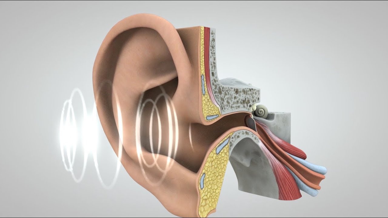
Image taken from the YouTube channel Neuroscientifically Challenged , from the video titled 2-Minute Neuroscience: The Cochlea .
Where Are the Receptors for Hearing Located? The Astonishing Answer
The human ear is a remarkable organ, responsible for converting sound waves into signals that the brain can interpret as music, speech, and all other sounds we experience. The key to this process lies in specialized cells called hearing receptors. The answer to the question "where are the receptors for hearing located" is both precise and fascinating, involving intricate anatomical structures within the inner ear.
Unveiling the Inner Ear
To understand where the hearing receptors reside, we must first journey to the inner ear. This complex structure is situated deep within the temporal bone of the skull and houses both the auditory system (responsible for hearing) and the vestibular system (responsible for balance). The inner ear consists of several key components, but the crucial area for our discussion is the cochlea.
The Cochlea: A Snail-Shaped Structure
The cochlea is a spiral-shaped, fluid-filled structure resembling a snail shell. It's within this seemingly small space that the magic of sound transduction occurs. The cochlea’s primary function is to convert the mechanical vibrations from sound waves into electrical signals that can be transmitted to the brain. It does this with the help of our hearing receptors.
The Organ of Corti: Home to Hearing Receptors
Now, we arrive at the heart of the matter: the Organ of Corti. Located within the cochlea, the Organ of Corti is the sensory organ responsible for hearing. Specifically, it houses the specialized receptor cells known as hair cells. Therefore, the receptors for hearing are located within the Organ of Corti, inside the cochlea of the inner ear.
Hair Cells: The Actual Hearing Receptors
Hair cells are the actual sensory receptors. They are so named because they have tiny, hair-like projections called stereocilia extending from their surfaces. These stereocilia are crucial for detecting movement within the fluid of the cochlea.
Types of Hair Cells
There are two main types of hair cells within the Organ of Corti:
- Inner Hair Cells (IHCs): These are the primary sensory receptors. They are responsible for transducing the mechanical vibrations into electrical signals that are sent to the auditory nerve. Approximately 3,500 inner hair cells exist in each cochlea.
- Outer Hair Cells (OHCs): These cells, numbering around 12,000 per cochlea, play a crucial role in amplifying and refining the incoming sound signal. They do this by changing their length, enhancing the movement of the basilar membrane (which will be explained later), which in turn improves the sensitivity and frequency selectivity of the inner hair cells.
The Basilar Membrane: A Vibrating Foundation
The Organ of Corti rests on the basilar membrane, a flexible structure that runs along the length of the cochlea. Different frequencies of sound cause different sections of the basilar membrane to vibrate. This vibration is crucial to stimulate the hair cells.
Frequency Mapping
The basilar membrane is tonotopically organized, meaning that different locations along its length respond maximally to different frequencies:
- Base (near the oval window): Sensitive to high frequencies.
- Apex (innermost part of the spiral): Sensitive to low frequencies.
This frequency mapping allows us to differentiate between various sounds.
A Step-by-Step Explanation of the Hearing Process
Let's recap the journey of sound and how it stimulates the hearing receptors:
- Sound Waves Enter: Sound waves enter the ear canal and cause the eardrum (tympanic membrane) to vibrate.
- Ossicles Amplify: These vibrations are amplified by three tiny bones in the middle ear (malleus, incus, and stapes).
- Oval Window Vibration: The stapes transmits the vibrations to the oval window, an opening to the inner ear.
- Cochlear Fluid Movement: The vibration of the oval window creates pressure waves in the fluid-filled cochlea.
- Basilar Membrane Vibration: These pressure waves cause the basilar membrane to vibrate.
- Hair Cell Stimulation: The vibration of the basilar membrane causes the stereocilia of the hair cells to bend.
- Electrical Signal Generation: Bending of the stereocilia opens ion channels, causing an electrical signal to be generated in the hair cells.
- Auditory Nerve Transmission: This electrical signal is transmitted via the auditory nerve to the brainstem and then to the auditory cortex in the brain, where it is interpreted as sound.
Factors Affecting Hearing Receptors
Several factors can damage hearing receptors, leading to hearing loss:
- Noise Exposure: Prolonged exposure to loud noise is a major cause of hair cell damage.
- Age: As we age, hair cells can gradually degenerate, leading to age-related hearing loss (presbycusis).
- Ototoxic Medications: Certain medications can be toxic to the inner ear and damage hair cells.
- Infections: Some infections can affect the inner ear and damage hair cells.
- Genetics: Genetic factors can also contribute to hearing loss.
Understanding the location and function of hearing receptors is crucial for appreciating the complexity and fragility of the auditory system. Protecting these sensitive cells is essential for maintaining healthy hearing throughout life.
Video: Hearing Receptors: The Shocking Location Revealed!
Hearing Receptors: Location FAQs
Here are some frequently asked questions about the surprising location of hearing receptors and how they work. Hopefully, this clarifies any lingering confusion!
So, where exactly are the receptors for hearing located?
The receptors for hearing are located within the inner ear, specifically in the cochlea. This coiled, snail-shaped structure houses the specialized hair cells that transduce sound vibrations into electrical signals.
Why is the inner ear considered a "shocking" location?
While the inner ear's role in hearing is well-known now, historically, understanding the intricate mechanisms within this tiny space was a significant scientific challenge. The complexity and delicacy of the inner ear receptors are still quite remarkable.
How do these receptors actually work?
Sound vibrations enter the ear and cause fluid in the cochlea to move. This movement bends the tiny hair cells inside. Bending these hair cells triggers the release of neurotransmitters, creating an electrical signal that travels to the brain.
What happens if these hearing receptors are damaged?
Damage to the hair cells in the cochlea can lead to hearing loss. This damage can be caused by loud noise, aging, certain medications, or genetic factors. Because the receptors for hearing, are so delicate, protecting your hearing is essential.