DNA Fragments: The Shocking Truth About Gel Electrophoresis!

Gel electrophoresis, a fundamental technique in molecular biology, relies heavily on the physical properties of DNA fragments. The size of DNA fragments significantly influences their mobility within the agarose gel matrix, a key component of the separation process. Scientists at institutions like the Broad Institute actively use this principle. This article examines how are dna fragments separated in gel electrophoresis, exploring the role of electric fields in driving these charged molecules through the gel and highlighting the importance of DNA ladders for accurate size determination.
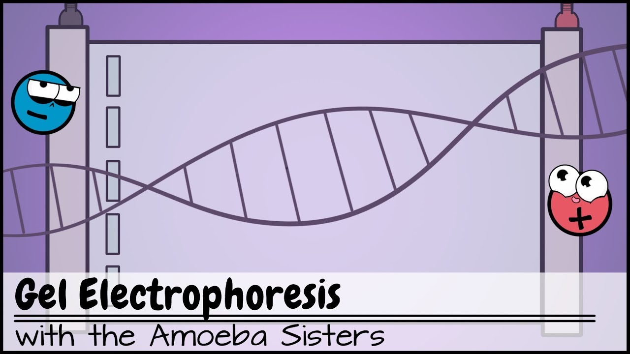
Image taken from the YouTube channel Amoeba Sisters , from the video titled Gel Electrophoresis .
Decoding the Separation: How DNA Fragments are Separated in Gel Electrophoresis
Gel electrophoresis is a cornerstone technique in molecular biology, allowing scientists to visualize and separate DNA fragments based on their size. This process relies on the physical properties of DNA and its interaction with an electric field within a gel matrix. This explanation details the mechanisms behind this separation.
The Foundation: The Electrophoresis Setup
Before delving into how DNA fragments are separated, understanding the basic components of gel electrophoresis is crucial.
The Gel Matrix: Our Molecular Sieve
The gel acts as a sieve, a porous material through which the DNA fragments must migrate. Two common types of gels are used:
- Agarose gels: Created from a polysaccharide derived from seaweed, agarose gels are typically used for separating larger DNA fragments (100 bp to 25 kb). The concentration of agarose determines the pore size, with higher concentrations creating smaller pores suitable for resolving smaller fragments.
- Polyacrylamide gels (PAGE): Formed from the polymerization of acrylamide and bis-acrylamide, PAGE gels offer higher resolution, particularly for separating smaller DNA fragments (5 bp to 500 bp) and proteins. The pore size can be precisely controlled by adjusting the acrylamide concentration and the ratio of acrylamide to bis-acrylamide.
The Electrophoresis Buffer: Conducting the Charge
The buffer solution serves two primary functions: it provides ions to carry the electric current and maintains the pH at a stable level, which is essential for DNA integrity. Common buffers include:
- TAE (Tris-acetate-EDTA): Cost-effective and suitable for most DNA electrophoresis applications.
- TBE (Tris-borate-EDTA): Offers higher buffering capacity than TAE and is preferred for resolving small DNA fragments.
- SB (Sodium Borate): Provides even better resolution for smaller fragments and generates less heat than TBE.
The Electrophoresis Chamber and Power Supply
The electrophoresis chamber holds the gel submerged in the buffer. Electrodes are connected to a power supply, creating an electric field across the gel. The power supply delivers a controlled voltage or current, driving the movement of the charged DNA molecules.
The Driving Force: Electrophoretic Mobility
The separation of DNA fragments in gel electrophoresis hinges on a fundamental principle: charged molecules migrate through an electric field.
DNA's Intrinsic Negative Charge
DNA possesses a consistent negative charge due to the phosphate groups in its sugar-phosphate backbone. This uniform charge-to-mass ratio is a key factor in gel electrophoresis separation. Because DNA is negatively charged, it will migrate towards the positive electrode (anode) when an electric field is applied.
The Impact of Fragment Size
The rate at which a DNA fragment migrates through the gel is inversely proportional to its size. Smaller fragments encounter less resistance within the gel matrix and move faster, traveling further towards the anode. Conversely, larger fragments experience greater resistance and migrate more slowly, remaining closer to the origin (the well where the DNA was loaded). This size-dependent migration is the foundation of DNA fragment separation.
The Mechanics of Separation: Navigating the Gel
The gel matrix plays a crucial role in differentiating DNA fragments based on size.
The "Sieving" Effect: A Molecular Obstacle Course
As DNA fragments migrate through the gel, the porous structure of the gel acts as a sieve. The pores create a network of obstacles that impede the movement of larger fragments more significantly than smaller ones.
| Fragment Size | Resistance in Gel | Migration Speed | Distance Traveled |
|---|---|---|---|
| Small | Low | High | Far |
| Large | High | Low | Short |
Factors Affecting Migration Rate
Several factors can influence the migration rate of DNA fragments:
- Gel concentration: Higher gel concentrations (e.g., more agarose or acrylamide) create smaller pores, increasing resistance and slowing migration.
- Voltage: Increasing the voltage increases the electric field strength, accelerating migration. However, excessively high voltages can cause the gel to overheat and melt or distort DNA bands.
- Buffer composition and ionic strength: The buffer's ionic strength influences the conductivity of the gel and the electric field strength.
- Temperature: Elevated temperatures can affect the gel's pore size and the DNA's mobility. Gels are often run in the cold room to help reduce this.
- DNA conformation: Supercoiled, linear, and open circular DNA fragments of the same size will migrate at different rates. This is more relevant for plasmid DNA analysis than for standard PCR product analysis.
Video: DNA Fragments: The Shocking Truth About Gel Electrophoresis!

FAQs: DNA Fragments and Gel Electrophoresis
Gel electrophoresis can seem complex, so we've compiled some frequently asked questions to help clarify how this crucial process works.
What exactly are DNA fragments, and why are they important?
DNA fragments are simply pieces of DNA that have been broken down or cut into smaller sizes. These fragments are crucial for analysis because they allow scientists to study specific regions of DNA, identify variations, and even create DNA fingerprints. Think of it like breaking a large book into chapters for easier study.
What is the "shocking truth" about gel electrophoresis?
The "shocking truth" refers to the fact that how are DNA fragments separated in gel electrophoresis is due to their negative charge being drawn to a positive electrode. The electric field pulls the negatively charged DNA fragments through a gel matrix, which acts like a sieve. Smaller fragments move faster and farther than larger ones.
What kind of gel is used for gel electrophoresis?
The most common type of gel is agarose, a natural polysaccharide derived from seaweed. Its pore size allows DNA fragments of various sizes to move through it. Polyacrylamide gels are also used, especially for separating smaller DNA fragments with very subtle size differences.
How does gel electrophoresis help in identifying individuals?
The how are DNA fragments separated in gel electrophoresis is by size is critical for DNA fingerprinting. By comparing the patterns of DNA fragments created from different individuals, scientists can determine if there is a match, which is useful in forensics, paternity testing, and other applications.